

Publication

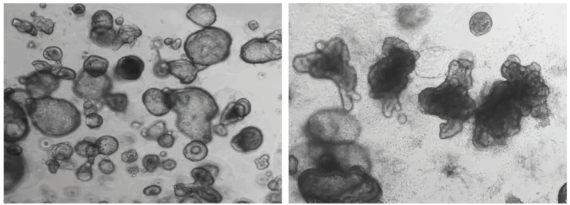
Intestinal cell morphology
The organoids initially form spheroid structures which over successive passages develop the crypt architecture characteristic of primary human intestinal organoids.
Figure 1. Typical intestinal morphology is observed in our iPSC-derived Intestinal organoids. Intestinal organoids grown encapsulated in matrigel in a 24 well plate.
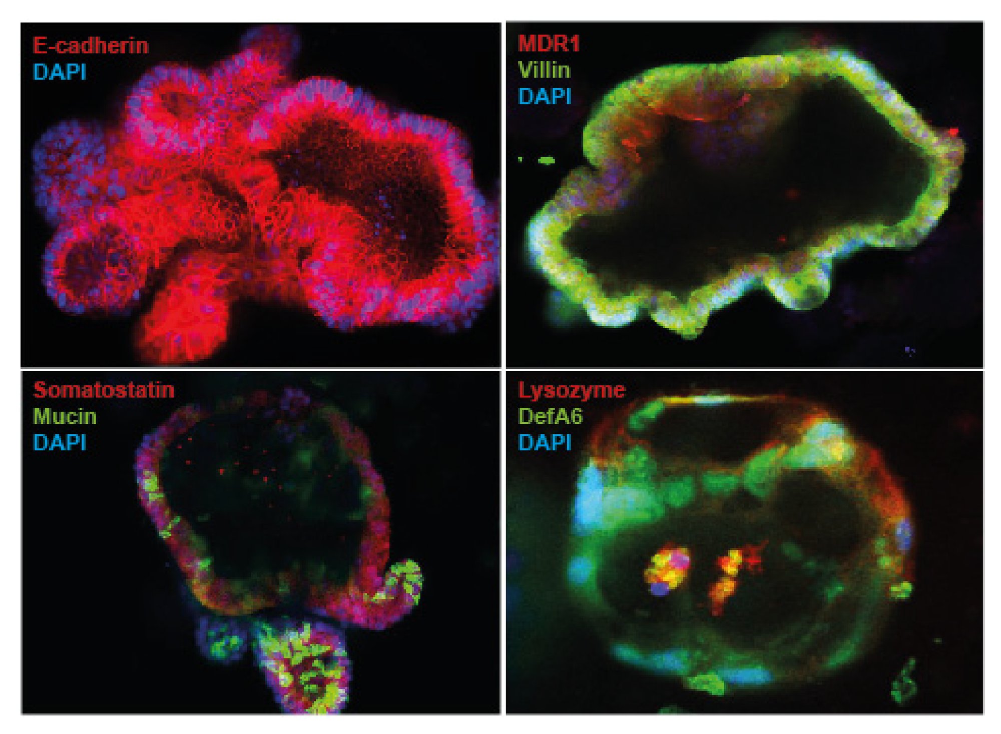
Key intestinal cell marker analysis
Immunocytochemistry analysis has demonstrated that the Intestinal organoids display a polarized epithelium and are composed of differentiated cell types with distinct morphologies. The organoids contain absorptive enterocytes as well as the major secretory lineage cell types including Paneth cells, goblet cells, and enteroendocrine cells.
Figure 2. Organoids display specific marker gene expression profiles. Organoids display positive staining of key intestinal cell markers including: epithelial cells (E-cadherin), enterocytes (villin), goblet cells (mucin), enteroendocrine (somatostatin), and Paneth cells (lysozyme).
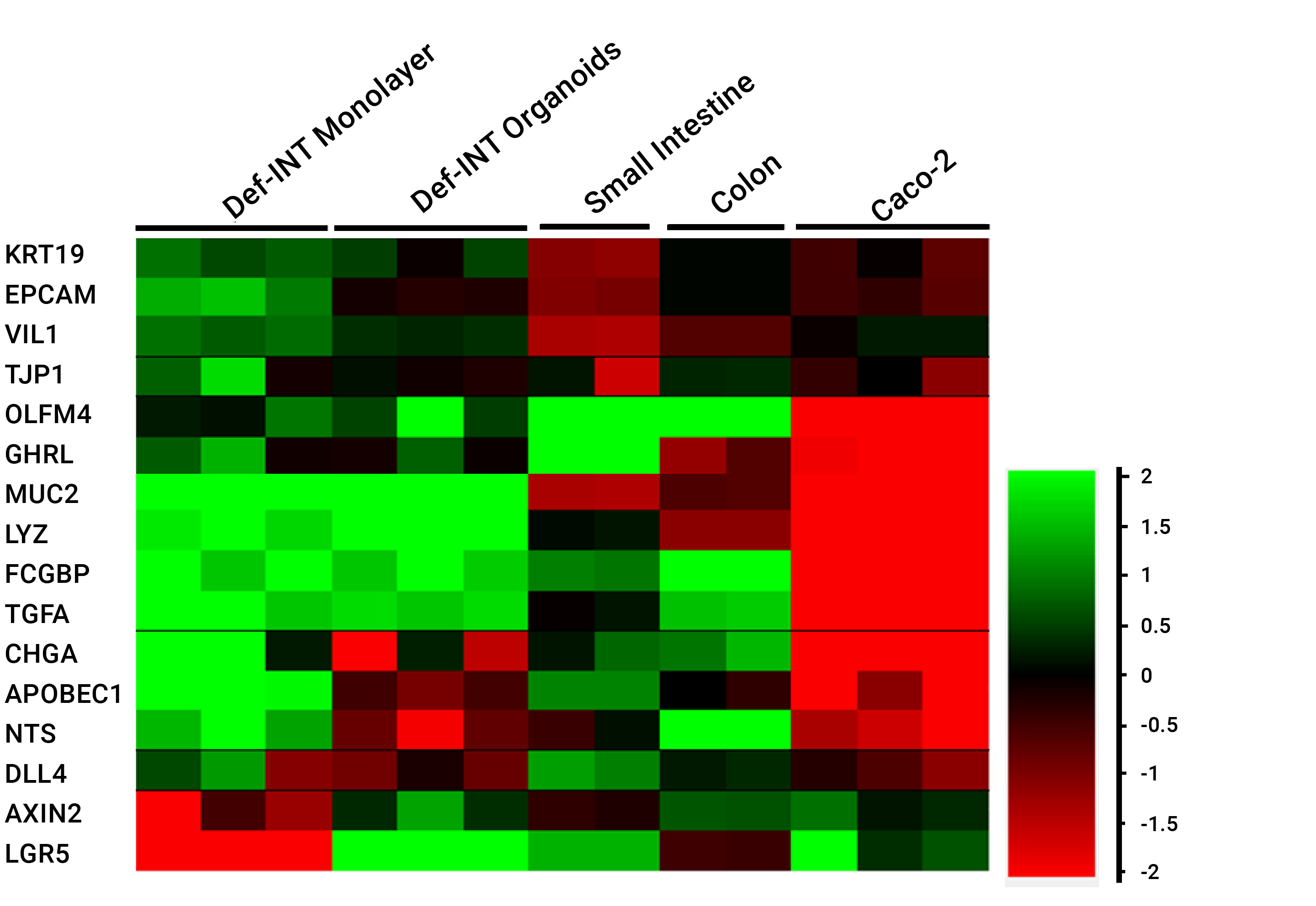
Our iPSC-derived Intestinal organoids have been demonstrated to display multiple key gut markers. Gene expression analysis shows key intestinal markers including intestinal stem cells (LGR5, Axin2), enterocytes (Vil1, EPCAM), goblet cells (MUC2, FCGBP), Paneth cells (LYZ, DLL4) and enteroendocrine cells (CHGA, GHRL) have similar expression levels in Intestinal organoids and the primary control relative to GAPDH.
Figure 3. iPSC-derived Intestinal organoids show gene expression profiles representative of intestinal stem cells, enterocytes, goblet cells, Paneth cells and enteroendocrine cells.
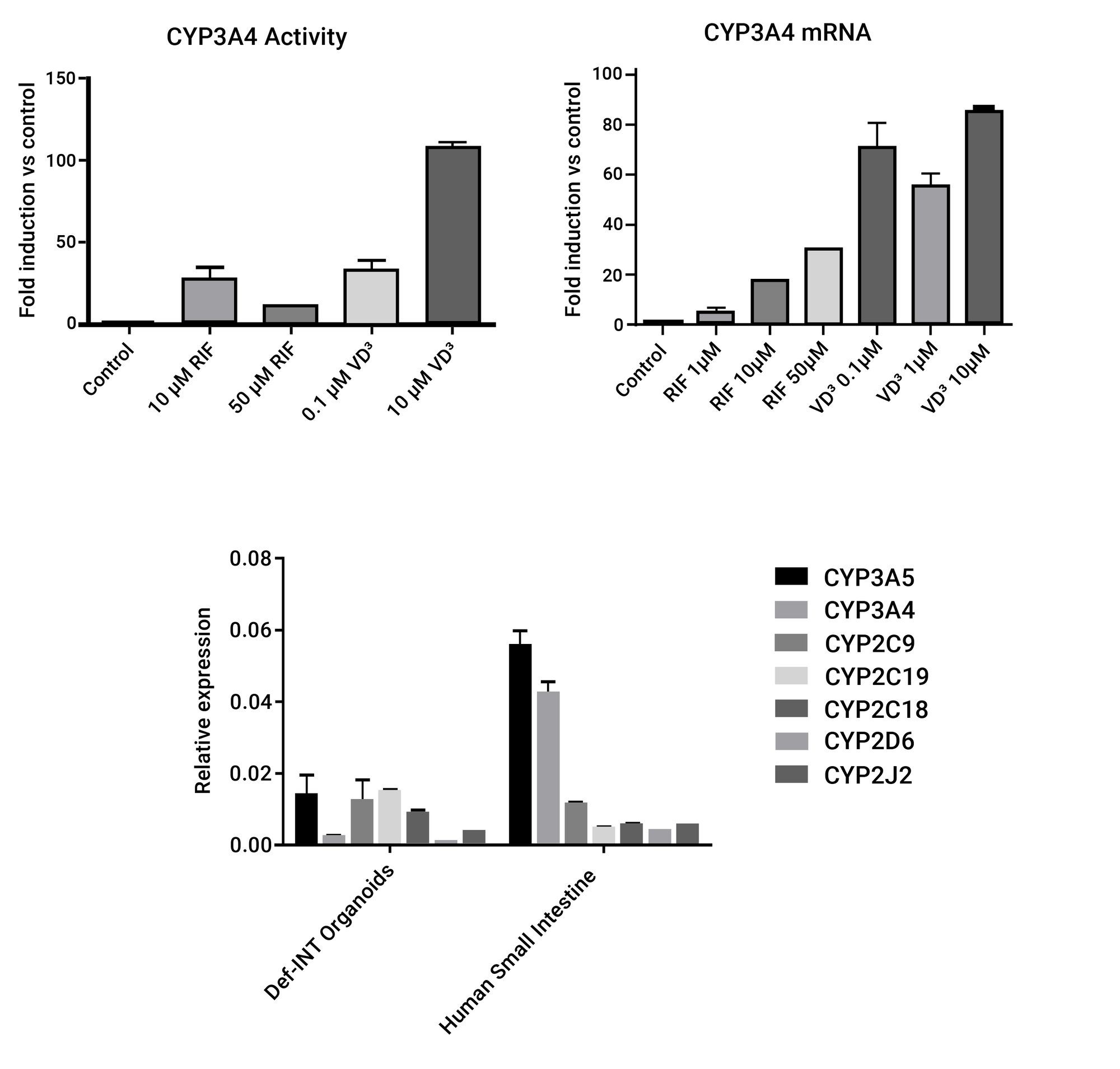
Phase I and Phase II metabolism
A range of Phase I and Phase II enzymes have been identified in the Intestinal organoids including CYP450, UGT, GST and CES. CYP3A4 is the highest CYP enzyme expressed in the human intestine and is involved in the metabolism of many drugs. Our Intestinal organoids display activity of CYP3A4 that can be induced upon incubation with RIF and VD3 at the mRNA and protein level. A range of other relevant isoforms of the CYP450 superfamily are also expressed in the organoids, including CYP3A5, CYP2C9, CYP2C19, CYP2J2 and CYP2D6.
Figure 4. CYP450 activity in the Intestinal organoids. A) CYP3A4 activity can be induced with different doses of RIF and VD3. B) CYP3A4 mRNA expression can be induced with RIF and VD3. C) Intestinal organoids express the major isoforms of the CYP450 family at similar levels to human small intestine.
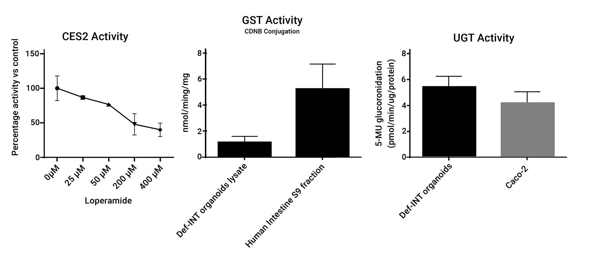
Other key enzymes for drug metabolism and detoxification are also active in the Intestinal organoids. The intestinal-specific isoform of Carboxilesterase (CES2) shows activity that can be inhibited by Loperamide. GST activity shown is at comparable levels of that found in S9 intestinal fractions and UGT activity is higher than that found in Caco-2 cells.
Figure 5. Activity of other Phase I and Phase II enzymes. A) CES2 activity present in the organoids. B) GST activity measured by conjugation of 2,4-Dinitrochlorobenzene (CDNB). C) UGT activity measured by glucuronidation of 4-Methylumbelliferone (4-MU).
Transporter expression and activity
The Intestinal organoids display gene expression of the key transporters involved in drug uptake and efflux, including MDR1 (ABCB1), BCRP (ABCG2), MRP2 (ABCC2), PEPT1 (SLC15A1) and OATP2B1 (SLCO2B1). Efflux transporters such as MDR1 and MRP2 can be localized in the apical side of the organoids. Activity of MDR1 has been shown by the ability of organoids to transport Rhodamine123, a MDR1 specific substrate, into the lumen. This activity can be drastically reduced with the competitive inhibitor Verapamil.
Figure 6. Transporter expression and activity in the organoids. A) Gene expression of the main intestinal transporters. B) MDR1 and MRP2 protein expression and localization. C) Rho123 assay showing MDR1 activity.

Let's work together